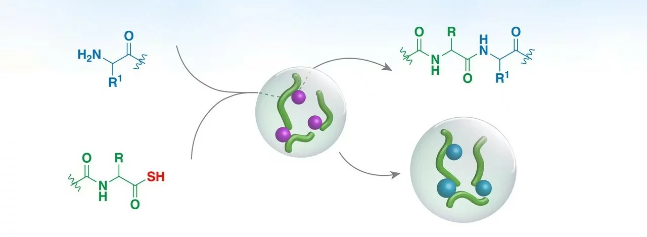
A peptide bond is the amide bond between the alpha-carboxyl group of one amino acid and the alpha-amino group of the next. It forms by condensation with loss of water, holds six atoms in a rigid plane, and hydrolyses so slowly in neutral water that the uncatalysed reaction takes hundreds of years. The chemistry below applies to the synthetic research peptides NuVion Health supplies as laboratory chemicals for in vitro research use only.
Formation by condensation
Joining two amino acids turns a carboxylic acid and an amine into an amide plus water. At neutral pH the equilibrium lies on the side of the free amino acids, so every route to a peptide bond first activates the carboxyl group, converting its hydroxyl into a better leaving group, and then lets the amine attack the activated carbonyl carbon.
The ribosome activates each amino acid as an aminoacyl-tRNA ester, charged by an aminoacyl-tRNA synthetase at the cost of ATP. In the peptidyl transferase centre, built from 23S ribosomal RNA, the alpha-amino group of the aminoacyl-tRNA in the A site attacks the ester carbonyl of the peptidyl-tRNA in the P site, and the growing chain is transferred onto the new residue. Ribosomal synthesis therefore runs from N-terminus to C-terminus.
Chemical synthesis activates the carboxyl group with a coupling reagent. Carbodiimides such as DIC, used with an additive such as OxymaPure, convert the acid into an active ester, and aminium reagents such as HBTU and HATU do the same through a benzotriazole ester. The free amine of the resin-bound chain then attacks the ester. Because every amino acid carries both an amine and a carboxyl group, the amine of the incoming residue is blocked with an Fmoc group and reactive side chains with acid-labile groups, so exactly one bond forms per coupling step. Solid-phase synthesis therefore runs from C-terminus to N-terminus, the opposite direction to the ribosome.
Geometry: why the bond is planar
The amide nitrogen carries a lone pair that delocalises into the carbonyl group. The C-N bond is about 1.33 Å long, between a single bond (1.47 Å) and a double bond (1.27 Å), and has roughly 40% double-bond character. Rotation about it costs about 80 kJ/mol, so at room temperature the bond is locked and the six atoms of the peptide unit (two alpha-carbons, the carbonyl carbon and oxygen, the nitrogen and its hydrogen) lie in one plane.
Two arrangements of that plane are possible. In the trans form (omega angle near 180°) the two alpha-carbons sit on opposite sides of the C-N bond; in the cis form they sit on the same side and clash. Cis bonds occur mainly before proline, where the ring removes the steric advantage of trans. Slow cis-trans isomerisation at X-Pro bonds is why proline-containing peptides can show doubled signals in NMR spectra.
Because the peptide unit is rigid, the flexibility of a chain comes from the two single bonds on either side of each alpha-carbon, described by the angles phi and psi. Steric clashes limit the accessible combinations to the regions of the Ramachandran plot that correspond to the alpha-helix, the beta-strand and polyproline II. Regular patterns of hydrogen bonds between the N-H of one peptide unit and the C=O of another define secondary structure.
Spectroscopic signature
The delocalised amide group absorbs ultraviolet light strongly near 190 nm, with a tail past 220 nm. That is why analytical HPLC of peptides uses detection at 210 to 220 nm: every residue contributes a chromophore, so a peptide without tryptophan or tyrosine is still seen and area-percent purity can be read from the trace. Circular dichroism in the far UV reads the same backbone; an alpha-helix gives minima near 208 and 222 nm and a disordered chain a single minimum near 200 nm.
Stability and hydrolysis
The resonance that makes the bond planar also makes it kinetically stable. Breaking an internal peptide bond quickly needs strong acid (6 M hydrochloric acid at 110 °C for 24 hours is standard for amino acid analysis), strong base, or a protease. Proteases accelerate the reaction by holding the bond in a strained geometry, activating a nucleophile and stabilising the tetrahedral intermediate. Sequence context matters: Asp-Pro bonds cleave in mild acid, Asn-Gly and Asp-Gly motifs rearrange through a succinimide intermediate to give deamidation and isoaspartate, and the terminal bonds are exposed to exopeptidases.
A lyophilised peptide has almost no water available for hydrolysis and is kept sealed, dry and away from light, refrigerated per the product documentation. Once reconstituted with bacteriostatic water it is a solution in which any contaminating protease can act, so it is refrigerated and used within the period on the documentation; the reconstitution calculator gives the concentration for a chosen volume. Synthetic peptides are often modified to slow enzymatic cleavage: a D-amino acid, such as the D-phenylalanine at position 7 in Melanotan 1, is not recognised by proteases, and N-terminal acetylation and C-terminal amidation block exopeptidases.
Isopeptide and related bonds
A bond formed between a side-chain carboxyl or amine and another residue is an isopeptide bond. Glutathione is linked through the gamma-carboxyl of glutamate, and ubiquitin attaches to proteins through the epsilon-amine of lysine. Synthetic cyclic peptides use the same chemistry: Melanotan 2 is closed by a lactam between aspartate and lysine side chains, formed on the resin after selective deprotection of those two residues. The other common ring closure, a disulfide between two cysteines as in oxytocin, is a different bond type.
Frequently asked questions
Why does the peptide bond not rotate freely?
The nitrogen lone pair is delocalised into the carbonyl, giving the C-N bond partial double-bond character. Rotation would break that conjugation at a cost of about 80 kJ/mol, so the bond stays planar.
Which peptide bonds are most likely to break during storage?
In dry lyophilised powder, essentially none. In solution, bonds next to aspartate are the most acid-sensitive, Asn-Gly and Asp-Gly motifs can rearrange through a succinimide, and any protease contamination cleaves at its own sites.
What is the difference between a peptide bond and an isopeptide bond?
A peptide bond joins the alpha-carboxyl of one residue to the alpha-amino of the next along the backbone. An isopeptide bond involves a side-chain carboxyl or amine, as in glutathione or a lactam-bridged cyclic peptide.
Browse the NuVion research peptide catalogue.
View productsResearch use only. This product is a laboratory chemical supplied for in vitro research. It is not included in the Australian Register of Therapeutic Goods and has not been assessed by the Therapeutic Goods Administration for quality, safety or efficacy. It is not for human or veterinary use, and nothing on this page is a representation about therapeutic use.